Imagine water flowing through a pipe. When the pipe is wide and flexible, the water flows easily and smoothly. But if the pipe is old, narrow, or clogged, the water has to flow faster and with more pressure to get through. The water doesn’t have as much space to move, and it faces more resistance. Our arteries work in a similar way. When they are flexible, like a wide, smooth pipe, blood flows easily and at a normal speed. But when the arteries become stiff, like a narrow or clogged pipe, blood has to move faster to get through, which is a sign of arterial stiffness.
Pulse wave velocity (PWV) is a key parameter used to assess arterial stiffness, or the flexibility of the arterial walls. When arteries are less flexible, blood moves through them faster, so a high PWV usually means the presence of arterial stiffness. This matters because stiff arteries make the heart work harder and are linked to a higher chance of cardiovascular diseases. To measure PWV accurately, various Pulse Wave Velocity Measurement Methods are used, providing doctors with valuable insights into a person’s cardiovascular system condition [1].
As discussed, there are various Pulse Wave Velocity Measurement Methods, each offering a unique approach to assessing the health of our arteries. Some methods are straightforward, while others employ advanced technology, but all serve to provide critical insights into the condition of our blood vessels. These methods range from simple, direct techniques to more complex, indirect approaches, including cutting-edge imaging and estimation technologies. In the following sections, we will explore the primary methods used to measure Pulse Wave Velocity (PWV), providing a clear overview of each technique along with the tools and steps involved in their application [2].
- Oscillometric
- Tonometry
- Photoplethysmograph (PPG)
- Doppler Ultrasound
- Magnetic Resonance Imaging (MRI)
- Pulse Transit Time (PTT)
Oscillometric Method for Pulse Wave Velocity Measurement
Oscillometric blood pressure measurement is a widely used method in automated blood pressure devices. This technique estimates blood pressure (BP) by detecting subtle changes, or oscillations, in arterial pressure during the inflation and deflation of a cuff.
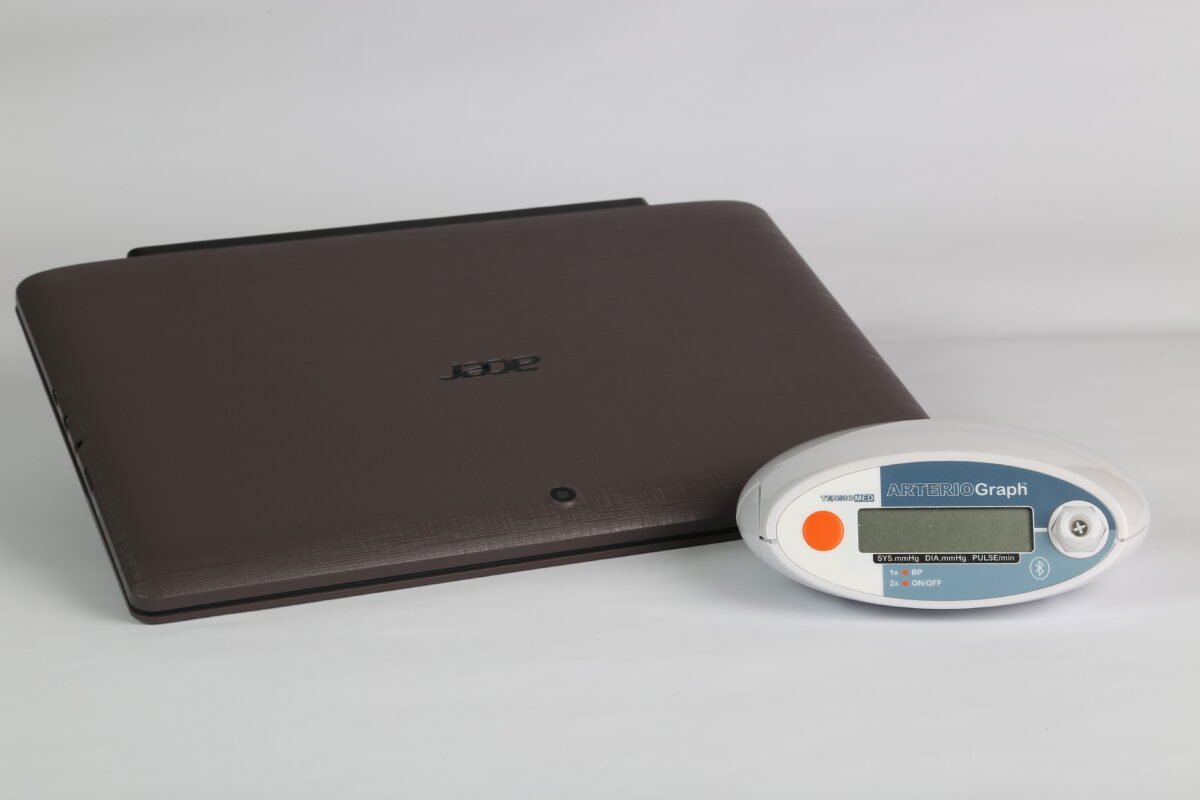
Various algorithms and methods are employed across different devices and manufacturers. However, one of the most robust validated approaches, verified through invasive techniques, is the supra-systolic pressure method used in Arteriograph, developed by Prof. Miklós Illyés. This method stands out for its precision and reliability [3].

Step-by-Step Process of the Supra-Systolic Pressure Method:
Initial Blood Pressure Measurement:
The oscillometric device begins by performing a standard blood pressure measurement. This provides the baseline systolic (SBP) and diastolic blood pressure (DBP) values for the individual.
Example: If the subject’s SBP is 130 mmHg and DBP is 80 mmHg, these values are recorded for subsequent steps.
Cuff Inflation to Supra-Systolic Pressure:
The device inflates the cuff to a pressure slightly above the subject’s systolic blood pressure. Specifically, it inflates to 35-40 mmHg above the SBP.
Example: For an SBP of 130 mmHg, the cuff inflates to 165-170 mmHg.
Pressure Wave Detection:
At this supra-systolic pressure level, the device measures pressure waveforms by detecting both:
- Direct pressure peaks: These are pressure waves traveling from the heart to the peripheral artery.
- Reflected pressure peaks: These waves reflect back from the peripheral vasculature.
The, the device determines the return time of the pressure waves (i.e., the time it takes for the wave to travel to a reflection point and return). This is a critical value for calculating PWV.
PWV Calculation:
The formula used for Pulse Wave Velocity is:
PWV= Return Time/Distance
The distance between the measurement points, such as the jugulum (base of the neck) and the symphysis (front of the pelvis), represents the approximate length of the aorta. This value is often estimated based on the individual’s height and body structure or measured directly in advanced setup.
Return Time: The time recorded by the device for the pressure wave to return.
Example Calculation:
Distance: Assume the measured aorta length is 0.5 meter (50 cm).
Return Time: The device records a return time of 0.050 seconds.
PWV = 0.5/0.05 = 10 m/s
Tonometry Method for Pulse Wave Velocity Measurement
Tonometry, originally developed for ophthalmology (Eye care), has evolved into a vital technique in cardiovascular health, allowing for a non-invasive assessment of arterial stiffness. This method employs precise sensors placed over major arteries, such as the carotid or femoral, to measure the velocity at which the pulse wave, generated by each heartbeat, travels through the arteries [4].
Preparation:
The individual lies down comfortably, relaxed, while the test is conducted.
Placement of Sensors:
Sensitive sensors, known as tonometers, are positioned on the skin over major arteries: commonly the carotid artery (located in the neck), the femoral artery (located in the groin), or sometimes at the wrist.
These sensors detect the pulse wave—the pressure wave produced by the heartbeat—as it moves along the artery.
How Tonometry Measures Pulse Wave Velocity
The tonometer records the time it takes for the pressure wave to travel between two points within the artery.
Device using the direct tonometry method: SphygmoCor (Cardiex Medical).
Photoplethysmograph Method for Pulse Wave Velocity Measurement
A Photoplethysmograph (PPG) works by shining light onto the skin and measuring how much light is absorbed or reflected. When the heart beats, blood flow in the arteries changes, which affects the amount of light absorbed. By tracking these changes in light, the PPG can detect each pulse [5]. For arterial stiffness, PPG looks at how fast the pulse wave travels through the arteries. In stiffer arteries, the wave travels faster, while in more flexible arteries, it moves more slowly. This speed (pulse wave velocity) gives insights into the artery health—higher speed usually means stiffer, less healthy arteries [6].
Preparation:
Where the Sensor Goes:
A PPG sensor is gently attached to a body part where blood flow is easy to detect, like a fingertip. Think of it as similar to the small clip doctors use on your finger to check oxygen levels.
Adding a Second Sensor (Optional):
If needed, a second PPG sensor may be placed somewhere else, such as on a toe or the opposite hand. This helps compare blood flow at two different spots in the body.
Using an ECG Sensor Instead:
Instead of using a second PPG sensor, a heart-monitoring sticker (ECG sensor) might be placed on the chest to track the heartbeat directly.
Measuring Distance:
The doctor or technician measures the distance between the sensors, or between a sensor and the heart. They use something simple, like a ruler or tape measure, to ensure the exact distance is recorded.
How Photoplethysmography Measures Pulse Wave Velocity
Recording the Pulse Transit Time:
For a single-sensor setup (PPG + ECG):
The time delay between the R-peak of the ECG signal (indicating the heartbeat) and the start of the pulse wave in the PPG signal is recorded.
For a dual-sensor setup (PPG at two locations):
The time difference between the arrival of the pulse wave at the first and second PPG sensors is recorded.
Device using the Photoplethysmographmethod: Withings ScanWatch
Doppler Ultrasound Method for Pulse Wave Velocity Measurement
The Doppler Effect is what happens when the pitch (how high or low the sound is) of a sound changes because of motion. Imagine you’re standing on the sidewalk, and an ambulance is driving toward you with its siren on:
- As the ambulance gets closer, the pitch of the siren sounds higher. This is because the sound waves are compressed as the ambulance moves toward you, so the sound reaches you more quickly.
- As the ambulance moves away, the pitch of the siren sounds lower. This happens because the sound waves are stretched out as the ambulance moves away, making the sound reach you more slowly.
This same idea is used in Doppler ultrasound in medicine. In Doppler ultrasound, high-frequency sound waves are sent into the body. These sound waves bounce off moving objects like red blood cells in blood vessels. When the blood cells are moving toward the ultrasound device, the sound waves get compressed (higher frequency), and when they move away, the sound waves get stretched (lower frequency) [7].

Preparation:
Where the probes Go:
A Doppler ultrasound probe is placed on the skin above the carotid artery (in the neck) and the femoral artery (in the groin). These arteries are chosen because they are easily accessible and provide valuable information about the heart’s function and the health of the blood vessels.
Using an ECG Sensor:
To synchronize the measurements with the heartbeat, an ECG sensor is placed on the chest. This sensor records the R-wave of the heartbeat, which helps the technician measure the exact timing of the pulse wave relative to the heart’s electrical signals.
Measuring Distance:
The technician measures the distance between the carotid artery and the femoral artery. This can be done using a simple measuring tape or the ultrasound machine, which provides the distance between the two locations where the measurements are taken.
How Doppler Ultrasound Measures Pulse Wave Velocity
The technician uses Doppler ultrasound to record the pulse wave in both the carotid artery and the femoral artery while synchronizing the measurements with the ECG signal.
The R-wave of the ECG marks the heartbeat.
The technician measures the time delay between the R-wave and the foot of the Doppler waveform, which is the point where the pulse wave starts in the arteries.
This time delay is the Pulse Transit Time (PTT), which is the time it takes for the pulse wave to travel between the carotid and femoral arteries [8].
MRI Method for Pulse Wave Velocity Measurement
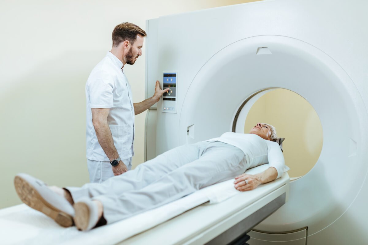
MRI, which stands for Magnetic Resonance Imaging, is a medical imaging technique that produces highly detailed pictures of the inside of the body. It is widely used by doctors to examine organs, tissues, and other structures without the need for an incision.
Pulse Wave Velocity (PWV) can be measured using several MRI techniques, which vary in complexity, accuracy, and imaging time:
2D Phase Contrast (2D PC) MRI
Two points along the aorta are selected (e.g., ascending and descending aorta).
MRI captures blood flow waveforms at these points.
The time difference (delay) between waveforms and the distance between points are used to calculate PWV.
Advantages: Simple and quick to perform.
4D Phase Contrast (4D PC) MRI
A 3D image of the entire aorta is captured over time.
Blood flow information is extracted from multiple locations along the vessel.
PWV is calculated using flow data from the entire vessel.
Advantages: Provides a comprehensive view of blood flow and minimizes errors from slice placement. It also provides additional information like pressure gradients and wall stress.
Disadvantages: Takes longer to perform (10-20 minutes) and has lower time resolution compared to 2D PC.
Fourier Velocity Encoded (FVE) MRI
A straight segment of the aorta is selected.
A narrow beam of MRI captures high-speed flow data along this segment.
Velocity waveforms are analyzed to calculate PWV.
Advantages: Very high time resolution (~3.5 ms), allowing detailed regional and global PWV measurements.
Disadvantages: Not suitable for curved or branching vessels, which are common in older or diseased arteries [9].

Pulse Transit Time Method for Pulse Wave Velocity Measurement
The PWV can be calculated using Pulse Transit Time (PTT), which is the time it takes for the pulse wave to travel between two points in the arterial system. To compute PWV, the distance between the two measurement points (D) is divided by the PTT:
PWV = D/PTT
The PTT itself can be measured noninvasively by obtaining the time difference between cardiac signals (usually the ECG) and peripheral pulse signals (typically obtained via PPG (Photoplethysmogram) sensors [10].
The ECG is used to detect the electrical signal from the heart, especially the R-wave, which marks the moment when the heart’s ventricles contract and pumps blood. The PPG sensor, typically placed on the fingertip, detects the arrival of the pulse at a more peripheral part of the body. The time difference between the R-wave (from the ECG) and the arrival of the pulse at the PPG sensor is recorded as Pulse Transit Time (PTT) [11].
Advantages and disadvantages of PPT Method:
One of the main advantages of measuring PWV based on PTT mrthod is that it is non-invasive. No needles or surgical procedures are required, and the measurements can be taken by wearing a heart monitor (ECG) and a PPG sensor on the skin. Continuous monitoring of blood pressure and arterial stiffness can be achieved, which is useful for preventing cardiovascular diseases. However, PTT measurements can be affected by motion, incorrect sensor placement, and even body temperature, leading to potential incorrect readings. Additionally, while PTT provides information about the timing of the pulse wave, it does not directly measure blood pressure, so it cannot fully replace traditional blood pressure measurements.
Suggested Pulse Wave Velocity Measurement method and device:
One of the easiest, most practical, and accurate ways to measure pulse wave velocity is by using the oscillometric method with the Arteriograph device. This device is simple, non-invasive, and quick, making it easy to use for both doctors and patients. The Arteriograph provides clear and reliable results by measuring arterial stiffness parameters, making it an excellent choice for accurate and convenient measurements.


