


The health of blood vessels is essential for overall well-being. Properly functioning vessels regulate blood flow, control inflammation, and maintain balance in the circulatory system [1]. Blood vessels are composed of several layers, with the innermost layer—the endothelium—playing a central role in their function. Endothelial Function Measurement allows these cells’ performance to be assessed, providing early insight into cardiovascular health. This article focuses on endothelial function and how it can be measured to detect potential problems before symptoms appear.
What Is endothelium?
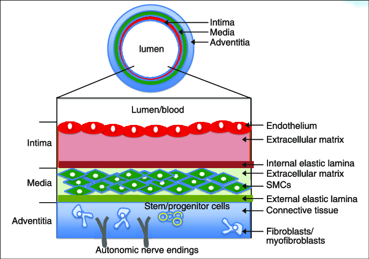
Blood vessels are composed of three layers, each with a specific role in maintaining circulatory health.
- The innermost layer, the tunica intima, is thin but essential. It is lined by endothelial cells, which regulate blood flow, vessel tone, inflammation, and repair processes. Changes in the endothelium can be detected long before symptoms appear.
- The tunica media, the thickest layer in arteries, is made of smooth muscle cells and elastic fibers. Strength and flexibility are provided, allowing vessels to expand and contract with each heartbeat.
- The tunica adventitia, the outer layer, provides structural support and nutrients. It is composed of connective tissue and contains nerves and small blood vessels that supply the larger vessel.
Among these layers, the endothelium—part of the tunica intima—is the most important. Even as a single cell layer, it controls blood flow, protects the vessel, and detects early problems. Measuring endothelial function can reveal cardiovascular risks before symptoms appear and guide preventive strategies [2].
What is endothelial Function and Why It Matters?
Endothelial function is how the inner lining of our blood vessels—the endothelium—keeps blood flowing smoothly, prevents clots, and protects the vessels from damage. Though it is just a thin layer of cells, it performs many vital tasks: controlling blood flow, reducing inflammation, and regulating the growth of the muscles in vessel walls. Two key molecules, nitric oxide (NO) and prostacyclin, play central roles in these processes [3].
- Nitric oxide (NO) is made from a substance called L-arginine. It acts like a natural relaxant for blood vessels, helping them widen so blood can flow smoothly. NO also acts as a protector, stopping inflammation, preventing clots, and keeping vessels in good shape. Blood flow itself and signals like bradykinin, adenosine, and VEGF tell the endothelium when to produce NO. When NO levels drop, other backup molecules help keep small vessels open, ensuring blood keeps moving.
- Prostacyclin is another important substance made by the endothelium. It relaxes blood vessels, prevents clotting, and shields vessel walls, but it works differently from NO. Together, NO and prostacyclin keep vessels flexible, open, and safe, making sure tissues get enough oxygen.
However, many cardiovascular risk factors—including obesity, inactivity, high cholesterol, high blood pressure, diabetes, smoking, and aging—can upset this balance. They increase oxidative stress, reduce NO availability, and shift the endothelium from a healthy, protective state to a dysfunctional one. Because of this, how well the endothelium dilates vessels is a key measure of overall vascular health. Poor endothelial function predicts long-term risks such as heart attacks, strokes, and high blood pressure. Keeping the endothelium healthy is therefore crucial for preventing cardiovascular disease and protecting your blood vessels [4].
How Endothelial Function Can Be Measured!
Endothelial function can be measured using invasive or noninvasive techniques [5]. Each method looks at different aspects of vascular health. For early disease detection, the ideal test should be noninvasive, reliable, reproducible, affordable, and easy to perform.
Invasive Methods:
- Intra-arterial infusion of acetylcholine (ACh) or endothelin: A small tube delivers these chemicals directly into an artery. Tools like strain gauges or high-resolution ultrasound measure changes in blood flow and vessel size.
- Intravascular ultrasound (IVUS): A tiny ultrasound probe is inserted into the coronary arteries. It takes detailed images of the vessel walls and any blockages.
Noninvasive Methods:
- Ultrasound flow-mediated dilation (FMD): A blood pressure cuff temporarily stops blood flow in the arm. When released, an ultrasound measures how much the artery widens, showing how well the endothelium works.
- Flow-mediated MRI: Uses MRI to see how vessels widen when blood flow increases. It evaluates endothelial function without touching the vessels.
- Pulse Wave Analysis (PWA): Measures arterial pressure waves using an oscillometric cuff. The augmentation index (AIX) is calculated from these waves. Lower AIX indicates flexible, healthy vessels. Higher AIX shows stiffer arteries and impaired endothelial function.
Flow-mediated dilation vs. Pulse Wave Analysis:
FMD is the gold standard for endothelial assessment. However, it is time-consuming, operator-dependent, and sensitive to many factors. PWA is easier, faster, and more practical. With modern technology, it provides reliable, noninvasive measurements of endothelial health using AIX. It is less operator-dependent and suitable for both clinical and research use.
Clinical Applications of Endothelial Function Measurement
Endothelial function measurement plays an important role in prevention, risk assessment, and patient care. Since endothelial dysfunction often appears before visible symptoms, it can be used as an early warning system. Detecting changes in vessel health at this stage allows prevention strategies—such as lifestyle improvements or medical treatment—to begin before serious problems, like heart attacks or strokes, develop.
It also improves risk assessment. Traditional tools, such as cholesterol levels or blood pressure, do not always give a complete picture. Measuring endothelial function provides direct insight into how healthy the blood vessels really are. This makes risk prediction more accurate and personalized for each patient.
Finally, endothelial function measurement supports patient care by tracking progress over time. It shows how well lifestyle changes, medications, or other treatments are working to restore vascular health. Noninvasive methods like Pulse Wave Analysis (PWA) make this practical in everyday clinical settings, offering quick, safe, and reliable evaluations.
Arteriograph and Endothelial Function Measurement
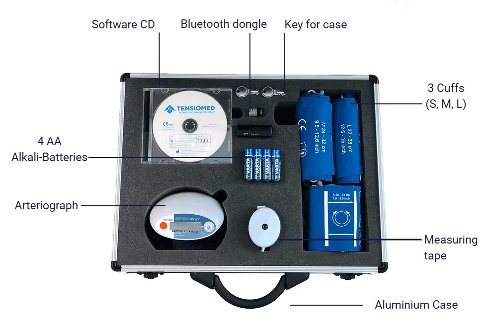
The Arteriograph is a noninvasive device that assesses arterial health using pulse wave analysis (PWA). The test is performed with a simple upper-arm cuff, much like a regular blood pressure measurement, making it quick, safe, and comfortable for patients.
One of the key parameters derived from PWA is the Augmentation Index (AIX). AIX reflects how flexible or stiff the arteries are and is widely recognized as one of the most important markers of endothelial function. A lower AIX indicates healthy, responsive vessels, while a higher AIX suggests stiffness and impaired endothelial performance.
In addition to AIX, the Arteriograph also measures pulse wave velocity (PWV), a strong indicator of arterial stiffness. Together, AIX and PWV provide a comprehensive picture of vascular health, combining information about both endothelial function and structural vessel changes.
Because it is noninvasive, reproducible, and fast, the Arteriograph makes endothelial function measurement practical in everyday clinical use. It supports prevention by identifying early dysfunction, improves risk assessment, and allows patient care to be tailored by monitoring how lifestyle changes or therapies affect vascular health.