

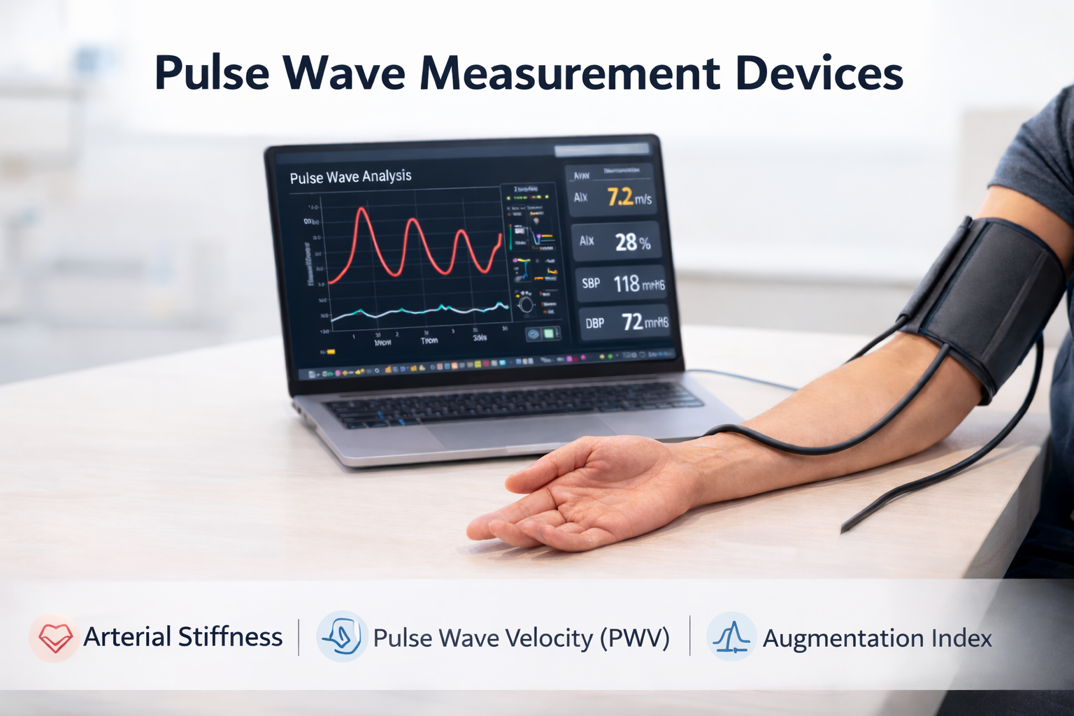
PWV measurement devices are important in both cardiovascular research and routine clinical practice, as pulse wave velocity (PWV) is widely used to assess arterial stiffness and overall vascular health.. Basically, Pulse Wave Velocity (PWV) is defined as the speed at which the pressure (pulse) wave generated by the heart propagates along the arterial tree. It reflects the stiffness of the arteries — stiffer arteries transmit the wave faster than more flexible ones — and is widely used as a non-invasive biomarker of arterial stiffness and cardiovascular risk [1].
What is the current status of PWV measurement?
According to the ESH Guideline 2023, PWV is an important measure of vascular aging, which naturally increases with age and is a focus of current research. Monitoring PWV can help doctors understand how stiff a patient’s arteries are and how their blood vessels are aging over time. In addition, studies have shown that reducing PWV is linked to better health outcomes, especially in patients with resistant hypertension or those on dialysis, making it a valuable tool for both risk assessment and treatment management [2].
Pulse Wave Velocity Measurement Principle
-
Distance = The actual length along a structure between two points, for example, from the root of the aorta to its bifurcation.
-
Transit time = It is the time it takes for the pulse wave to travel from one point, such as the root of the aorta, to another point, for example the abdominal aorta [3].
Invasive Measurement of Pulse Wave Velocity (PWV)
Non-invasive measurement methods of PWV
PWV can be measured using several non-invasive methods, which do not require surgery or entering the body. These techniques allow doctors and researchers to assess arterial stiffness safely and efficiently. The most common approaches include applanation tonometry, Doppler ultrasound, and oscillometric devices, each with its own advantages and typical uses.
Overview of PWV measurement devices
| Device Name | Type of Measured PWV | Measurement Method | On the Market Since |
| Arteriograph | Aortic PWV (PWVao, estimated) | Oscillometric based on supra-systolic method | 2004 |
| SphygmoCor | Carotid–Femoral PWV (cfPWV) | Applanation tonometry with ECG gating | 1990 |
| Complior | Carotid–Femoral PWV (cfPWV) | Tonometry / mechanotransducer sensors (point-to-point method) | 1984 -no longer produced |
| Vicorder | Carotid–Femoral PWV; Brachial–Ankle PWV | Oscillometric dual-cuff measurement | 2007 |
| Mobil-O-Graph | Estimated central/aortic PWV | Oscillometric cuff with transfer-function algorithm | 2010 |
| PulsePen | Carotid–Femoral PWV (cfPWV) | Applanation tonometry | 2004 |
Arteriograph:
The Arteriograph is an innovative device designed to make PWV measurement easy, fast, and reliable in everyday clinical practice. Key features include:
-
First patented device: Arteriograph is the first user-independent method for measuring PWV.
-
Simple and fast: Uses a single upper-arm suprasystolic cuff, making it suitable for daily clinical routine.
-
Clinically validated: Its measurements have been confirmed against invasive methods.
-
Supported by research: Several cardiovascular outcome studies demonstrate its reliability.
-
Direct measurement: Does not rely on a generalized transfer function (GTF), ensuring accurate assessment.

Complior
The Complior is a traditional method for measuring PWV that uses piezoelectric sensors. While reliable, it is relatively time-consuming, requires trained personnel, and is no longer available on the market. Key features include:
-
Established method: A well-known, traditional device for PWV measurement.
-
Requires training: Measurement needs skilled operators to ensure accuracy.
-
Clinically validated: PWVcf, SBPao, and AIXao measurements have been confirmed against invasive methods.
-
Supported by research: Multiple cardiovascular outcome studies demonstrate its reliability.
-
Direct measurement: Does not rely on a generalized transfer function (GTF), ensuring accurate assessment.
SphygmoCor Xcel
The SphygmoCor Xcel is a device designed for measuring arterial stiffness and pulse wave analysis. It combines oscillometric and applanation tonometry techniques, offering expensive but detailed hemodynamic assessment. Key features include:
-
Dual measurement methods: Uses a single upper-arm cuff for pulse wave analysis (PWA) and simultaneous carotid applanation tonometry + thigh oscillometry for cfPWV measurement.
-
Time and user dependent: PWV measurement can be relatively “time-consuming” and requires trained personnel.
-
Validated parameters: AIXao and SBPao measurements have been confirmed against invasive methods.
-
GTF usage: Uses a generalized transfer function (GTF) to estimate aortic pulse pressure from brachial artery waveforms, which has not been invasively validated for PWVcf.
-
Publication history: First described in the literature in 1990.
Vicorder
The Vicorder is a user-independent device designed for simple and simultaneous measurement of carotid-femoral PWV. Key features include:
-
Cuff-based method: Uses simultaneous carotid and femoral cuffs to measure PWVao.
-
User-independent: Easy to operate and suitable for routine clinical use.
-
Supported by research: At least one cardiovascular outcome study demonstrates its utility.
-
Limited validation: No invasive validation study is available for PWVcf, AIXao, or SBPao.

PulsePen
The PulsePen is an applanation tonometric device designed to measure aortic PWV. While seems accurate, it is user-dependent and requires trained personnel. Key features include:
-
Applanation tonometry with ECG gating: Measures PWVao using precise timing.
-
User-dependent: Measurement requires skilled operators and can be time-consuming.
-
Clinically validated: Aortic PWV has been confirmed against invasive methods.
-
No GTF used: Does not rely on a generalized transfer function (GTF).
-
Limited validation: No invasive validation studies are available for SBPao or AIXao.

Mobil-O-Graph
The Mobil-o-Graph is a user-independent device, although its accuracy may be limited and influenced by age, designed for simple and rapid PWV measurement using a single upper-arm diastolic cuff. Key features include::
-
Cuff-based method: Uses a single upper-arm diastolic cuff for measurement.
-
User-independent: Easy to operate and suitable for routine clinical use.
-
Limited PWV accuracy: PWVao measurements are considered unreliable due to the ARC Solver Algorithm. The measured PWV correlates with the patient’s age and systolic blood pressure (SBP), rather than true arterial stiffness.
-
Validated SBPao: Central systolic blood pressure (SBPao) has been confirmed against invasive methods.
-
GTF usage: Uses a non-validated generalized transfer function (GTF).
-
Limited validation: No invasive validation studies are available for AIXao.

Recommended PWV Measurement Devices
In summary, among the currently available non-invasive PWV measurement devices, the Arteriograph appears to be the most suitable choice due to its simplicity, speed, user-independence, and validated reliability. The SphygmoCor Xcel also provides accurate measurements and detailed hemodynamic assessment, making it a strong alternative, though its PWV measurement can be more time-consuming and user-dependent. Other devices, while valuable in research or specialized settings, have limitations in accuracy, validation, or ease of use for routine clinical practice