


Pulse Wave Velocity (PWV) is an indicator of cardiovascular health, specifically reflecting the elasticity of arteries and vessels. Measuring and monitoring PWV helps healthcare providers assess arterial health and identify individuals at risk of cardiovascular diseases. It also assists in making treatment decisions and interventions aimed at improving arterial elasticity and reducing cardiovascular risk.
This article will describe a few non-invasive devices for measuring pulse wave velocity, focusing on the most common ones.
1. Arteriograph
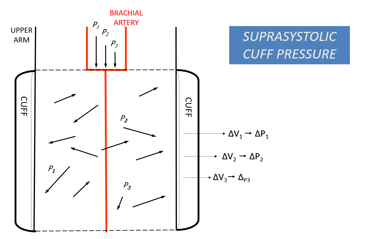
Method: Oscillometric method (Upper Arm Cuff Based on the Supra-Systolic Pressure)
Measured Parameters: 15 different parameters including Aortic Pulse Wave Velocity, Central Blood Pressure, and Aortic Augmentation Index.
How does the Arteriograph operate?
The device was developed by Prof. Ilyés, a Gynecologist and professor at the University of Pécs. It is based on an oscillometric method and uses an algorithm reliant on supra-systolic pressure, inflating the cuff on the upper arm to a pressure higher than the patient’s measured systolic blood pressure (approximately 35 to 40 mmHg higher). This completely closes off the brachial artery. When the brachial artery is fully occluded, any pulses (both direct and reflected waves) from the heart are transmitted to the tissues of the upper arm and then to the inflatable cuff. A very sensitive pressure sensor records these pulses, which are then displayed on the screen. The entire measurement process is automated and involves minimal human intervention.
Advantages:
- Invasive validation for Arterial Stiffness Parameters
- Highly reproducible
- Highly accurate
- User-friendly
- Quick
- Portable
- Painless
- Cost-effective
2. Complior
Method: Piezoelectric and Applanation Tonometry Methods
Measured Parameters: Normal Blood pressure values and Central Blood Pressure, Augmentation Index and Carotid-Femoral Pulse Wave Velocity.
How does Complior operate?
The device originates from France and uses a very complicated, user-dependent method. First, the patient should lie in a supine position while the operator measures peripheral pressure values using another machine and enters the data into the Complior software. Next, the distance between the carotid and femoral arteries is manually measured, which may involve human error, and then entered into the software. A sensor is then placed on the neck with gentle pressure. Another sensor must be placed by the operator on the femoral artery, requiring the patient to undress. The entire process is complex and demands skilled operators.
Advantage:
- Invasive Validation for Arterial Stiffness Parameters
- The delay in pulse transit time between two arterial sites is taken simultaneously using a ‘foot to foot’ waveform method which makes the measurement reliable.
- Requires operator skill.
- Carotid tonometry is challenging.
- The patient must undress and expose the groin.
- Technical errors may occur in obese patients.
- Distance measurement between the two arterial sites can be uncertain and approximate.
3. SphygmoCor XCEL
- Method: Piezoelectric and Applanation Tonometry Methods
- Measured Parameters: Normal Blood pressure values and Central Blood Pressure, Augmentation Index and Carotid-Femoral Pulse Wave Velocity.
How does SphygmoCor XCEL operate?
The device was developed in Australia. The assessment of Pulse Wave Velocity using the SphygmoCor XCEL requires the patient to be in a supine position. First, a cuff must be placed high up on the patient’s thigh, sometimes in direct contact with the skin. The operator then locates and marks the exact position of the carotid artery. Three separate distance measurements are required:
- From the suprasternal notch to the marked carotid artery.
- From the suprasternal notch to the top of the thigh cuff.
- From the femoral pulse to the top of the thigh cuff.
Additionally, normal blood pressure needs to be measured separately. The entire process is quite complicated and requires the operator’s skill.
Advantage:
- Invasive Validation for Arterial Stiffness Parameters
- PWA is available allowing assessment of the augmentation index and central BP through a transfer function application.
Depends on the operator’s skill.
A precise blood pressure calibration is needed for Pulse Wave Analysis (PWA).
ECG R-wave gating is used, but because the measurements are taken sequentially, the pulses recorded are not identical. This is due to the time gap of 2-5 minutes or more between the carotid and femoral measurements.
Conclusion:
Pulse wave velocity (PWV) measurement devices are crucial for assessing cardiovascular health by evaluating arterial elasticity and stiffness. Each device offers unique methodologies and benefits, including non-invasiveness, accuracy, and portability. However, challenges such as operator skill dependency and potential technical errors are notable for devices like SphygmoCor XCEL and Complior.
Among these devices, the Arteriograph stands out as a superior choice for measuring PWV. It is particularly notable for its accessibility in routine clinical settings, offering straightforward use compared to more complex methods.
Contact us to get answers to any questions you may have or to get a quote for the Arteriograph set.
Can you please provide a cost analysis for complete Arteriograph package
Dear Anthony Lawrence,
Many thanks for reading our article and for your inquiry.
We will contact you via email shortly.
Hello, Can you send an offer on the Arteriograph? We could be interested in one.
Best regards
Jakob
Hi Jakob Kaad,
Thank you for your interest! We’ll be in touch with you by email soon.
Best,
Hi, can you email me the price for the pulse wave velocity devices , along with the catalogue and interpretation method.
Dear Seowli Lim,
We have sent you an email with the requested documents.
Best regards,
Can you please email the price of the device
Dear Dr. Aitzaz Bin Sultan Rai,
We sent you an email and kindly request you to check your inbox at your earliest convenience.
Wishing you a great weekend!
Best regards,